
Nikon has announced the winners of its 47th annual Small World Photomicrography Competition. This year’s first-place prize was awarded to Jason Kirk for his striking image of a southern live oak leaf’s trichomes, stomata, and vessels.
The Nikon Small World competition was founded in 1974 to recognize excellence in photography through the microscope and is widely regarded as the leading forum for recognizing the art, proficiency, and photographic excellence involved in photomicrography. In 2011, the sister competition of Nikon Small World, Nikon Small World in Motion, was launched in response to technology advances allowing for recording movies or digital time-lapse photography through the microscope. The winners of that competition were announced on August 16.
This year, the competition received nearly 1,900 entries from 88 countries which were judged on originality, informational content, technical proficiency, and visual impact.
To capture his photo of an oak leaf, Kirk used various lighting techniques and design tools to create what Nikon’s judges determined to be a masterful example of the dynamic relationship between imaging technology and artistic creativity. Using a custom-made microscope system that combines color-filtered transmitted light with diffused reflected light, Jason captured around 200 individual images of the leaf and stacked them together to create the stunning image above.
Jason used both transmitted and reflected light on opposite sides of the leaf to highlight three vital structures. Prominently featured in white are the trichomes, which are fine outgrowths that protect a plant against extreme weather, microorganisms, and insects. In purple, Jason highlights the stomata, small pores that regulate the flow of gases in a plant. Colored in cyan are the vessels that transport water throughout the leaf. All three are essential to plant life.
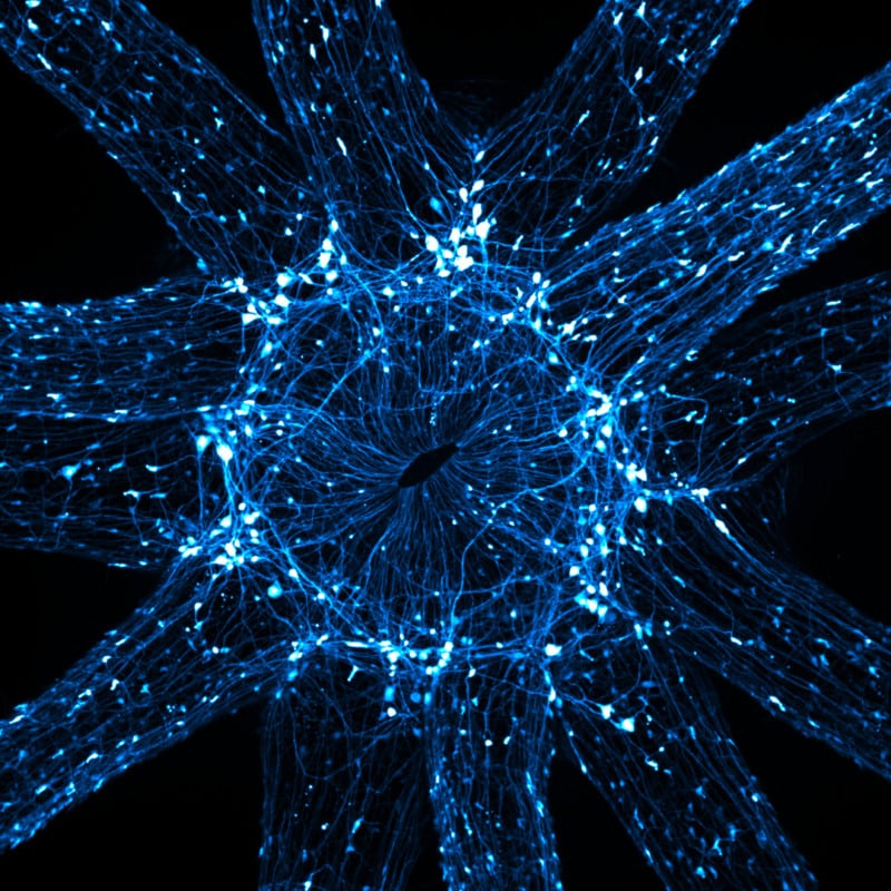
Second place was awarded to Esmeralda Paric for her image of a microfluidic device containing hundreds of thousands of networking neurons. The primary neurons were extracted and cultured, then seeded and transduced with a virus. The particular image shows two populations separated but bridged, with different viral treatments. It was maintained for 30 days, immunostained, and tiled imaged.
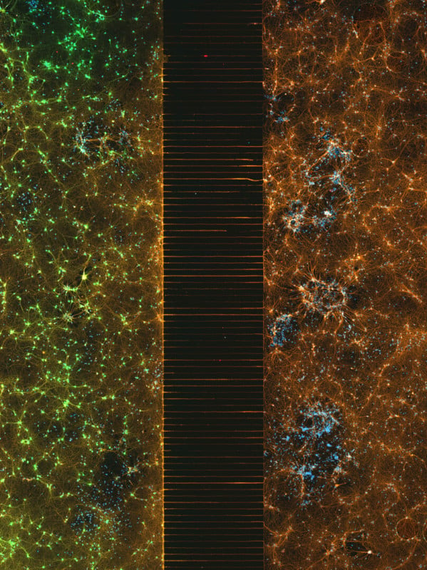
A microfluidic device containing 300k networking neurons in 2 isolated populations. Both sides were treated with a unique virus and bridged by axons. | Esmeralda Paric & Holly Stefen
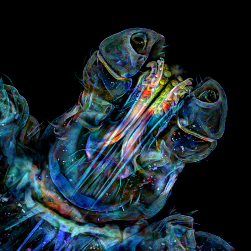
Third place went to Frank Reiser for his photo of a rear leg, claw, and respiratory trachea of a hog louse (Haematopinus suis).
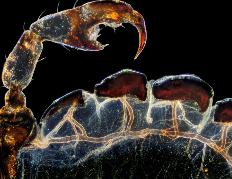
Rear leg, claw, and respiratory trachea of a louse (Haematopinus suis) | Frank Reiser
Below are the 17 other images — in order from fourth through 20th place — recognized in the top 20 photos submitted to the competition:
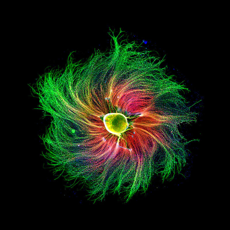
Sensory neuron from an embryonic rat | Paula Diaz
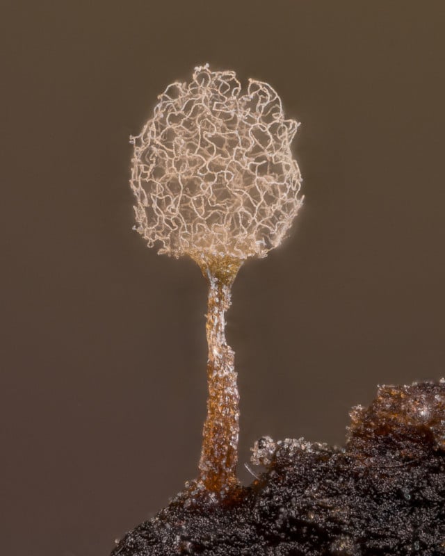
Slime mold (Arcyria pomiformis) | Alison Pollack
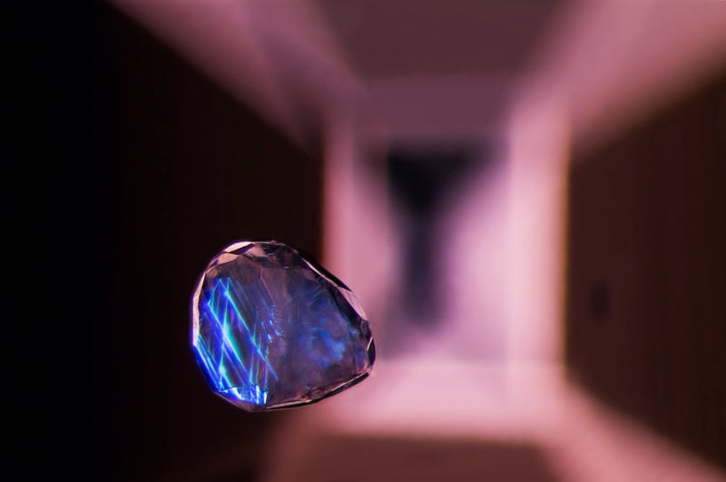
Calcite crystal inclusion suspended in a spinel gemstone | Billie Hughes
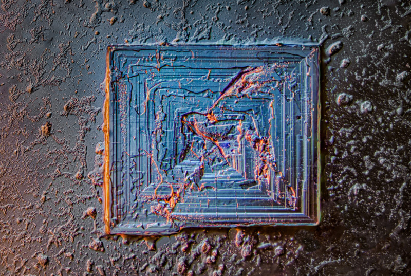
Table salt crystal | Saulius Gugis
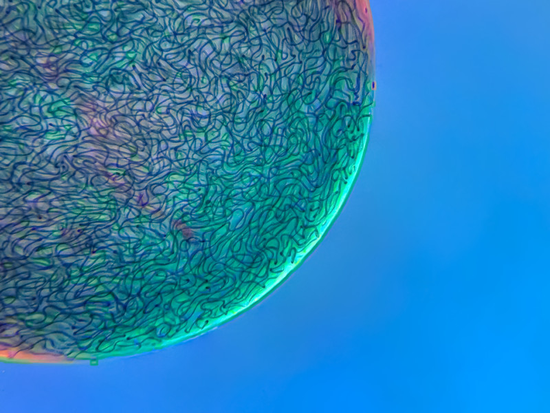
Filamentous strands of Nostoc cyanobacteria captured inside a gelatinous matrix | Martin Kaae Kristiansen
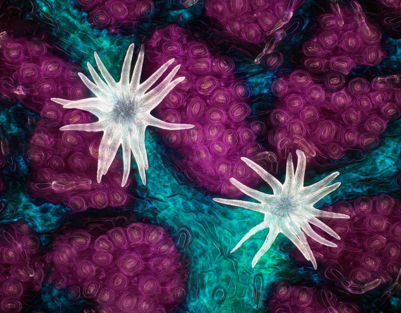
An in vivo snapshot of the neurons surrounding the mouth and tentacles of a juvenile starlet sea anemone (Nematostella vectensis) | Ruohan Zhong
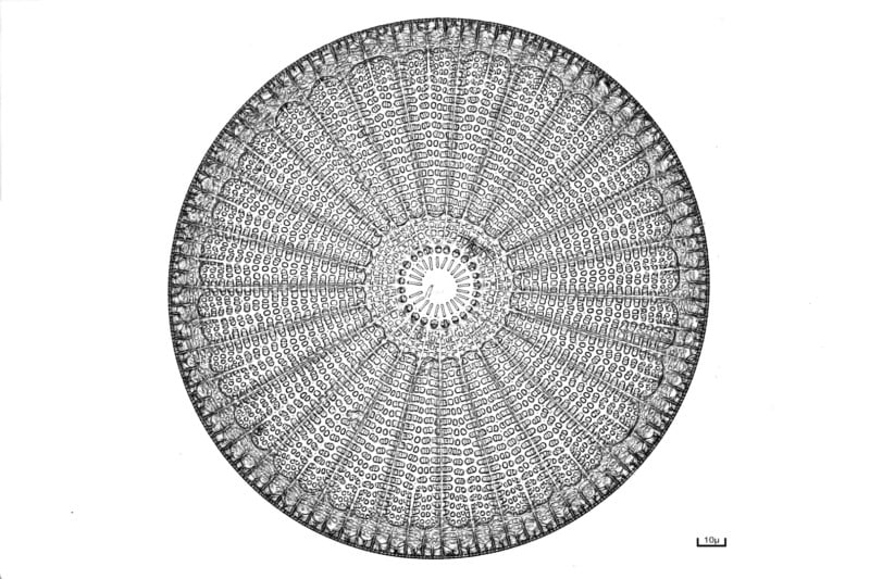
Diatom (Arachnoidiscus) | Bernard Allard
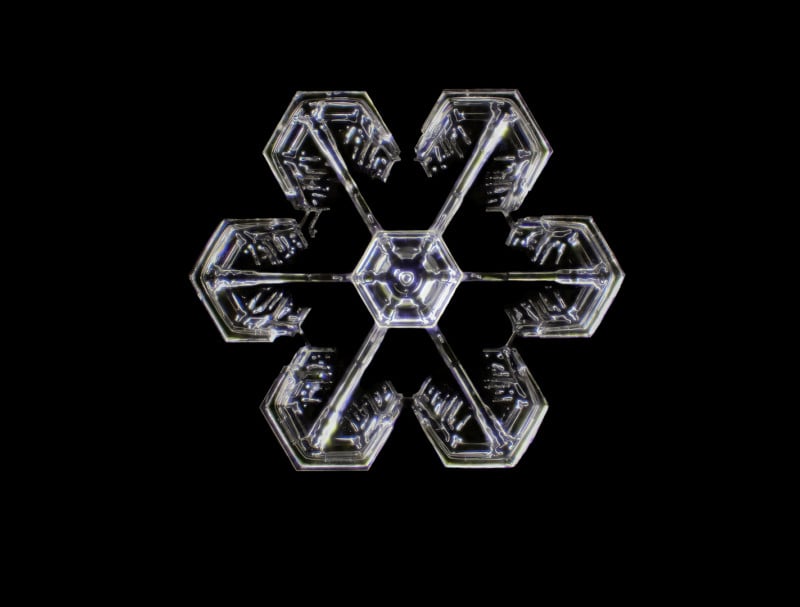
Snowflake | Dr. Joern N. Hopke
Cotton fabric with pollen grains | Dr. Felice Placenti
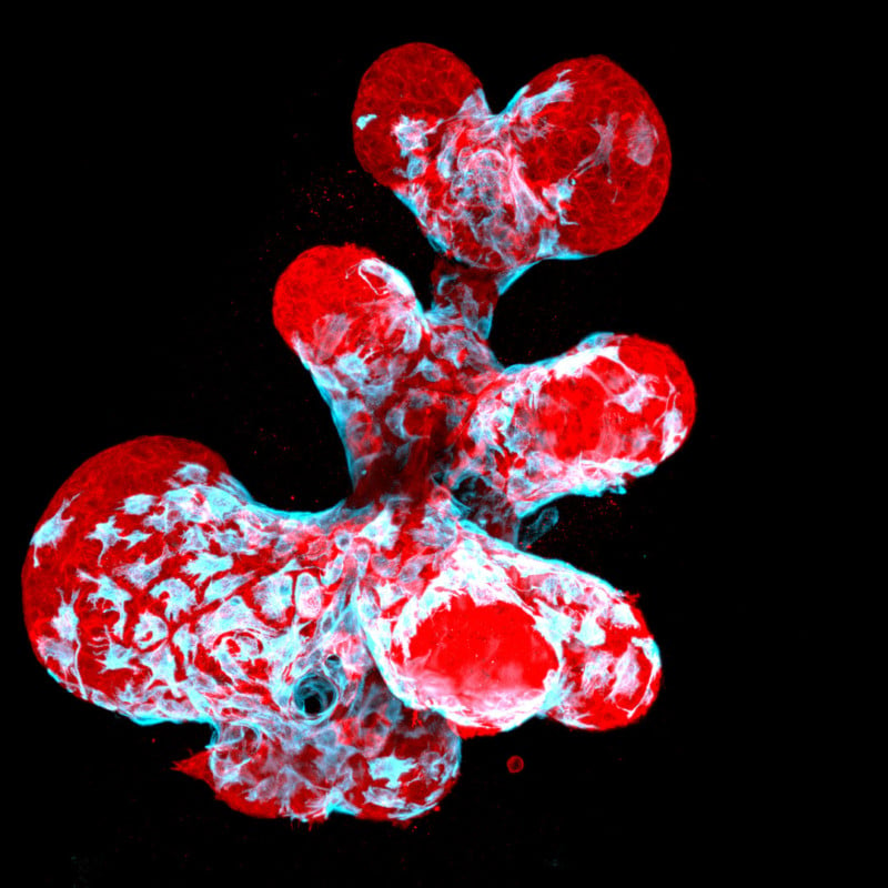
Breast organoid showing contractile myoepithelial cells (blue) crawling on secretory breast cells (red) | Jakub Sumbal
Vasculature of a mouse retina | Jason Kirk & Carlos P. Flores Suarez
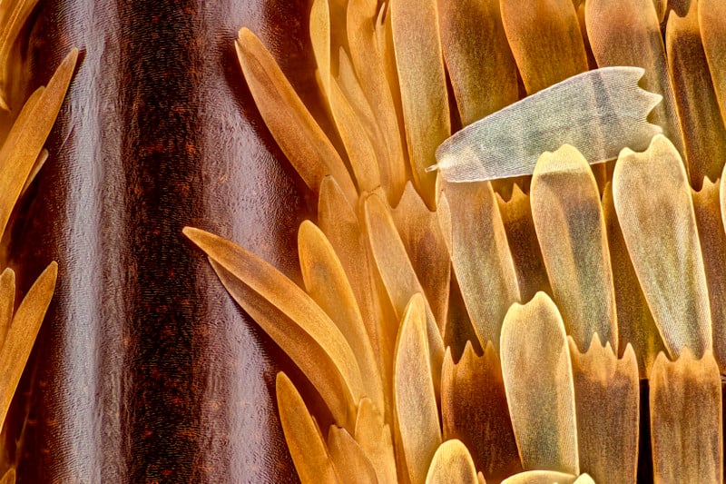
Vein and scales on a butterfly wing (Morpho didius) | Sébastien Malo
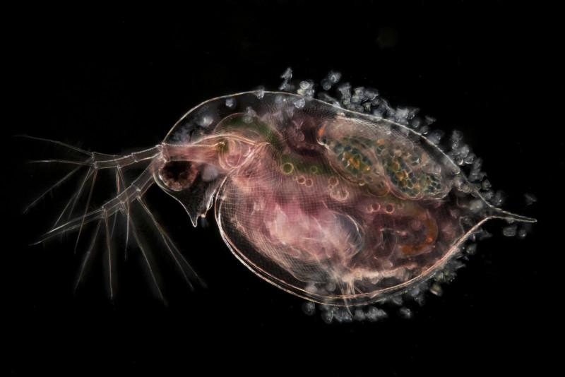
Water flea (Daphnia), carrying embryos and peritrichs | Jan van IJken
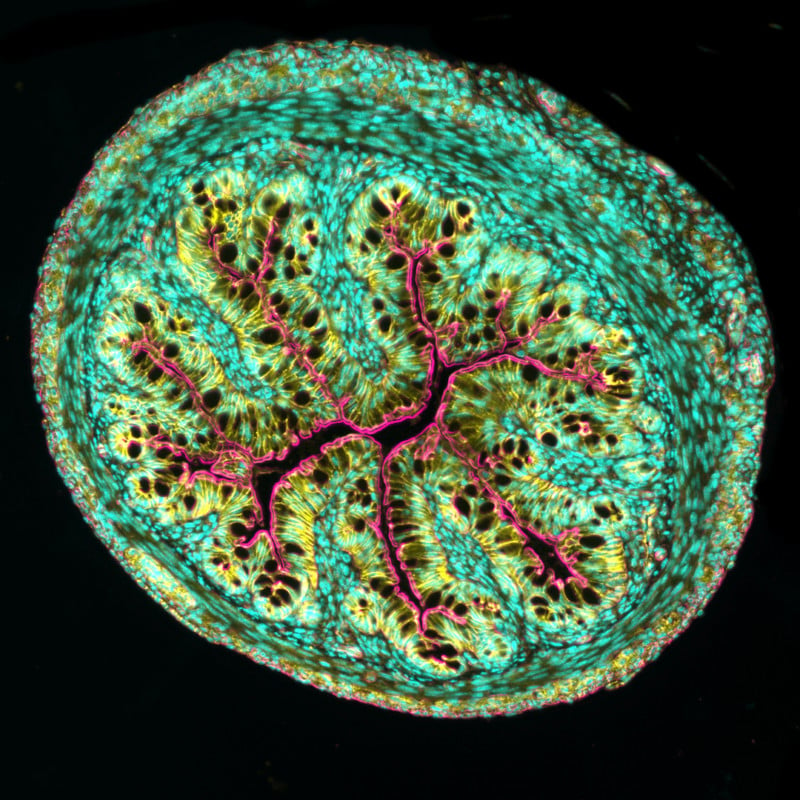
Cross section of mouse intestine | Dr. Amy Engevik
Head of a tick | Dr. Tong Zhang & Dr. Paul Stoodley
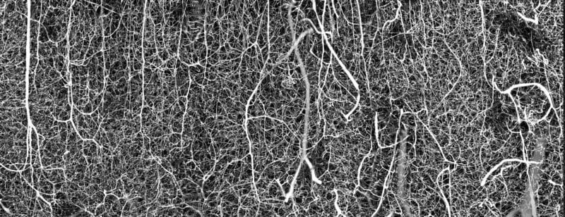
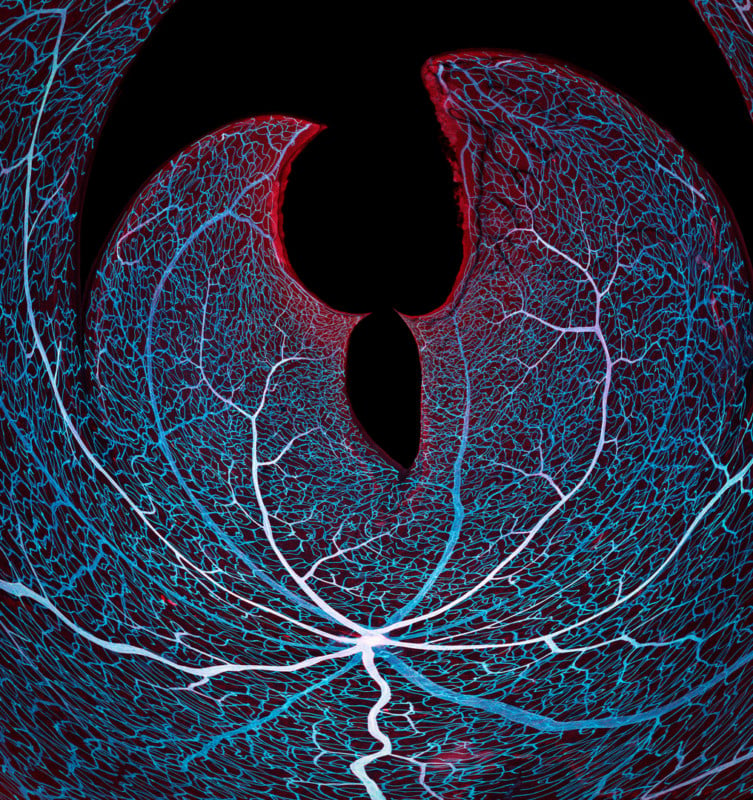
3D vasculature of an adult mouse brain (somatosensory cortex) | Dr. Andrea Tedeschi
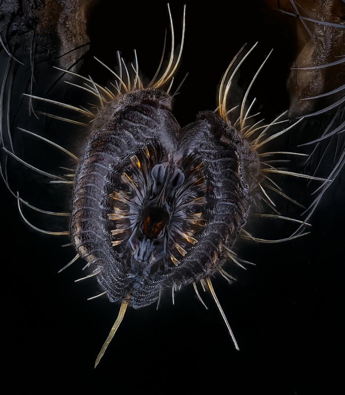
Proboscis of a housefly (Musca domestica) | Oliver Dum
A full-color calendar of all the winners is planned to be produced. To see all images from the competition that were recognized by the judges, visit the Nikon Small Worlds Competition website.
Image credits: Header image by Jason Kirk. All other photos individually credited and provided courtesy of Nikon Small World.
Continue reading...